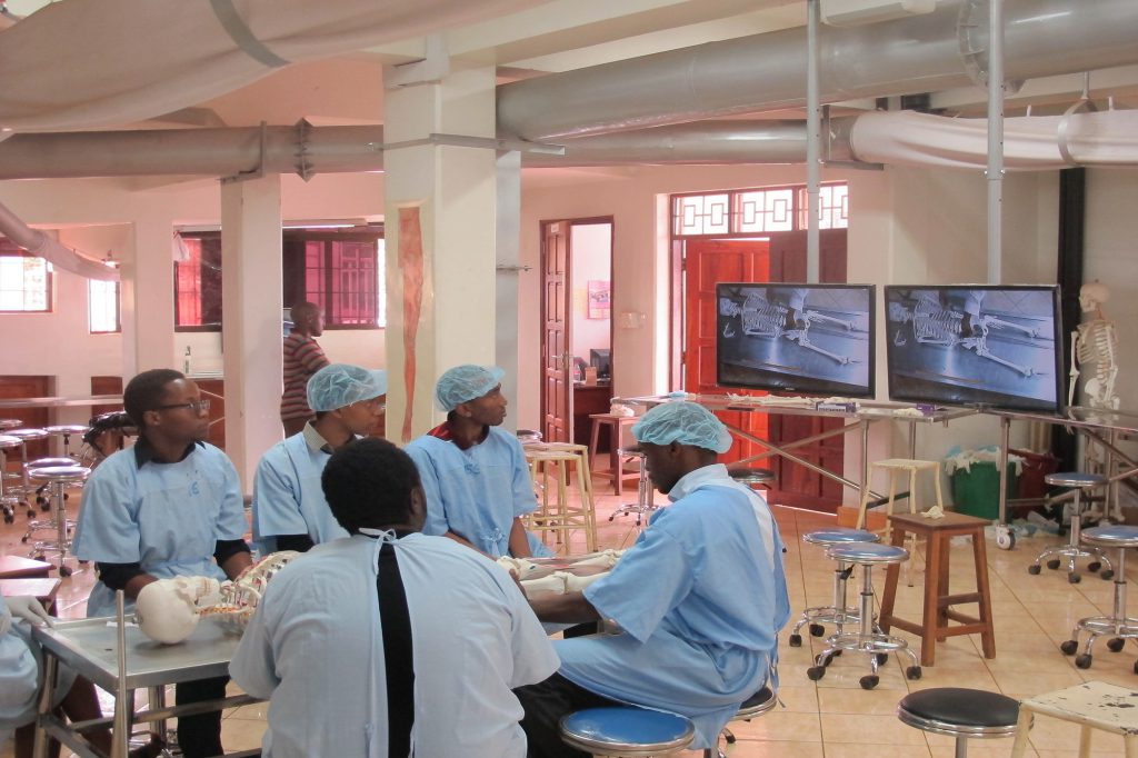
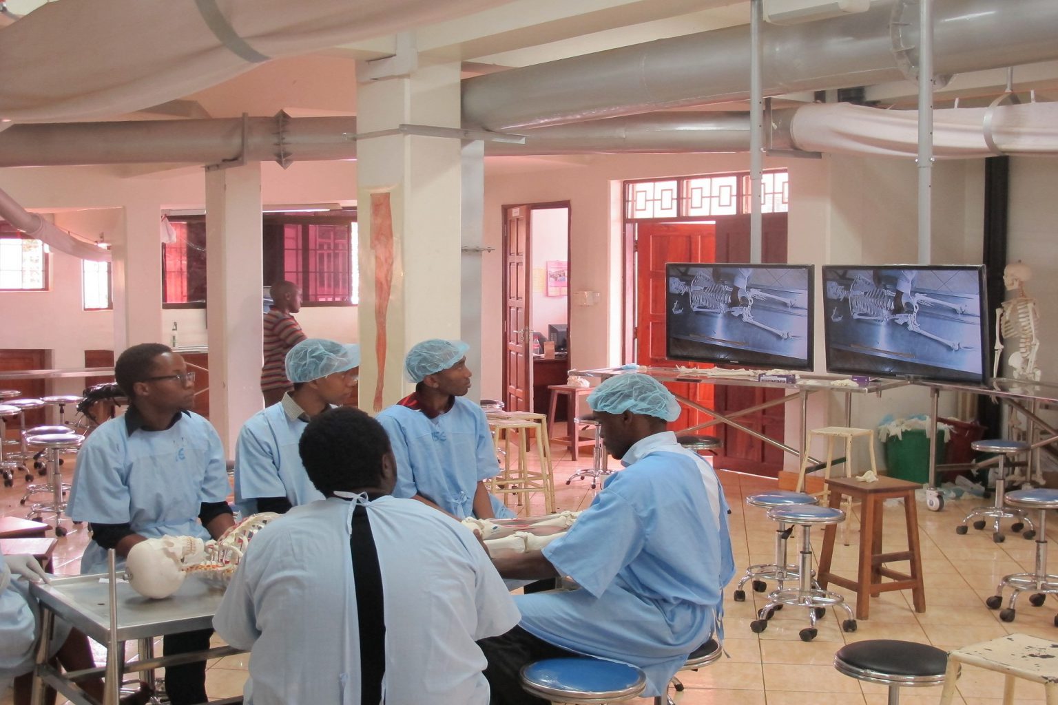
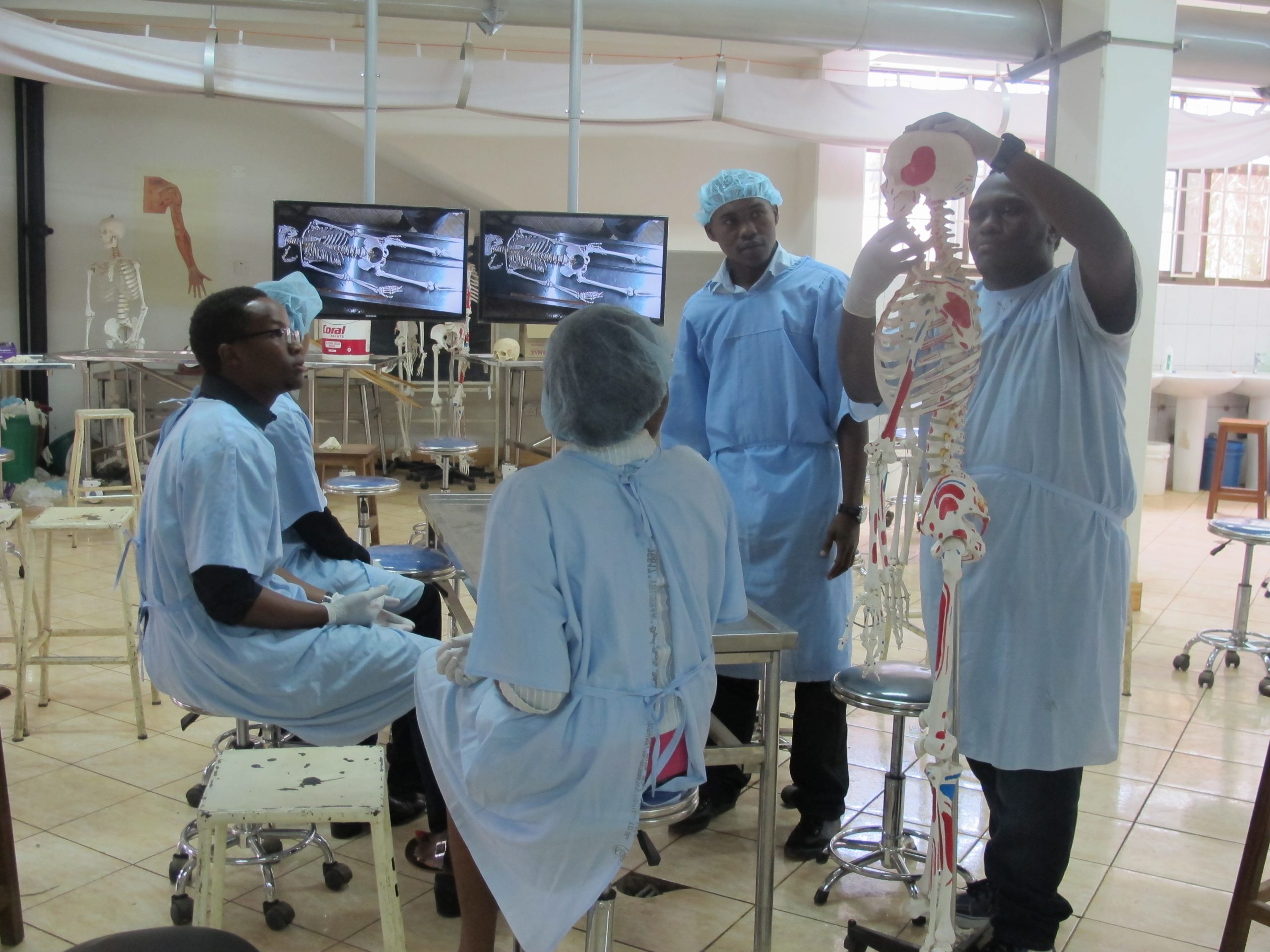
Anatomy laboratories in medical schools are places where students learn human clinical anatomy through the dissection of human cadavers. At KCMC, our anatomy laboratory accommodates 160 students at once and students are organized into dissection groups surrounding a table. Each group consisting of 12 to 16 students dissects one cadaver. In average, we have 10 to 13 dissection groups per year. Dissections, normally supervised by course instructors, are guided by a dissection manuals (Grants dissector 5th edn) read by one student in a group while other medical students dissect the cadaver. It is common for the students to find the task of following the manual and identifying the anatomical structures as a challenge. Therefore, the course instructors form an component to facilitate proper acquisition of the practical objectives. Due to limited course instructors, it was difficult to satisfy each group during a practical dissection and provide specialized attention to the unique challenges that each group faced. It was under this challenge that we received funding from Medical Education Partnership Initiative (MEPI) for installation of Digital Audio Visual and Recording (DAVR) system in our anatomy laboratory for enhanced students – instructor learning interactions and provide learning of anatomy behind the walls of the dissection laboratory.

The soon installed DAVR system is composed of 9 Samsung plasma flat screens, overhead speakers, overhead camera, two wireless microphones, video mixer and a recorder, allowing all the 160 medical year-one students to clearly follow the instructions from the course instructor. The students can now learn together as well as learn from the questions and responses posed by other students. Meanwhile the recorded DAVR practical clip is made available for viewing under the Learning Management System (LMS) after the sessions. This has greatly reduced the redundancy of information provided to individual groups and has objectively connected the instructor to the groups and vice versa; therefore, the new method has enhanced the achievement of the practical learning objectives at a shorter time in a large class setting. DAVR has improved students- instructor learning and teaching interactions during practical anatomy dissections and improved both their satisfactions and output. We anticipate, improved students performance in anatomy practical examinations, reduced practical dissection time and better instructor performance. In the future, we hope the practical DAVR clips will form an important complement for revising and learning clinical anatomy practical that can extend behind the walls of the dissection room.


